Anatomy of the Prostate Gland and Seminal Colículos of the Canine (Canis lupus familiaris)
Anatomy Physiology & Biochemistry International Journal Juniper Publishers

Abstract
The prostate gland and the seminal colliculi are within the classification of the genital organs of Canis lupus familiaris, although it can also be classified within the urogenital system of the canine. The glands are characterized by having the peculiarity of making secretions and these are regulated mainly by hormonal control and autonomic nervous system; On the other hand, in reference to the seminal colliculi, it is understood that it has an intimate relationship with the urethra and with other nearby anatomical structures. The anatomical links between the prostate gland and the seminal colliculi are quite close, since in both cases the urethra is involved. First; the urethra pierces the prostate and secondly; from the urethral crest a seminal colliculus is born. The prostate gland and the seminal colliculi have a fundamental link with the process of ejaculation, since the prostate makes a prostatic fluid, which helps the activation of the sperm and the seminal colliculus is connected to the vas deferens that carry the sperm from the epididymis to the urethra. Through this study a bibliographic review was made based on the anatomy of the prostate gland and seminal colins of the canine, information that has a pedagogical purpose involving general functions, structures, positions and anatomical relationships that can be analyzed by veterinary medicine students who are studying the subject of anatomy, specifically, the anatomy of Canis lupus familiaris.
Keywords: Prostate gland; Seminal colículos anatomy; General functions; Structures; Positions; Anatomical relationships; Urethra
Introduction
The prostate is a gland present in all male domestic mammals and is a mixed gland, since it is formed by serous and mucosal constituents [1]. The prostate is a globular gland that surrounds the neck of the bladder and the urethra during its union, it presents a body and two lobes or wolves (right and left) which discharge their contents to the urethra through prostatic ducts. The prostatic fluid plus the sperm coming from the testicles form the semen. The adnexal glands of the reproductive system in mammals present variations in relation to their location, number, size and shape, for the canines it is odd, and their size is similar to a chestnut [2]. During the life of the dog the development of the prostate can be divided into two fundamental stages, the first corresponds to the period of embryogenesis and immediate postnatal development and ends when the animal is 2-3 years old, the second stage consists of a phase of exponential hypertrophic development that is androgen dependent and ends when the animal is between 12 to 15 years of age [3]. Within the embryology or embryogenesis period, the prostate gland has an endodermal origin and arises from the pelvic urethral epithelium, there are a series of symmetric cavities and the prostatic tubules appear as solid epithelial projections of the urethra approximately in the sixth week of gestation.
The prostatic glandular epithelium develops from those endodermal cells; therefore, the mature prostate gland represents a fusion composed of mesenchymal, urethral and Wolffian duct tissue, these are all united within a common capsule that would be the prostate gland in its entirety [4]. The prostate within its functions adds prostatic secretions (prostatic fluid) to ejaculation to provide an optimal environment for the survival and mobility of sperm, it should be mentioned that this gland is present in all domestic species [5]. The urethra has two portions, a pre-prostatic and prostatic, which are summarized as the pelvic part (pars pelvina), in the dorsal region of its light the pre-prostatic part of the urethra presents a fold of the mucosa or urethral crest (crista urethralis) ending in the aforementioned seminal colliculus. In the dog and the cat, the body of the prostate is bulky, in the dog in addition to the vas deferens ampulla, the prostate is its only accessory genital gland, whereas in the cat there is the prostate, the ampulla of the vas deferens and find the pair formation of the bulbourethral gland [6].
Anatomical Description of the Prostate Gland of the Canine
The prostate gland (Figure 1) is the only accessory sex gland in the dog and unlike other domestic mammals, the canine lacks bulbourethral and vesicular glands [7]. This gland is relatively large and has a yellowish coloration [8]. The prostate is the most developed sexual gland that the dog possesses and has a body divided into two lobes, right and left that surround the urethra, it also has a longitudinal septum that separates the two lobes and the disseminated part of the prostate that is distributed dispersed around the urethra [9]. The prostate completely surrounds the neck of the bladder and the beginning of the urethra (Figure 2), the weight and normal size of the prostate are very variable, although this is usually located at the level of the pubic rim. It is a flattened gland on the back and rounded in the ventral direction and to the sides it is strongly encapsulated, from the ventral part of the capsule a longitudinal septum that reaches the prostatic portion of the pelvic urethra is detached, thus dividing partially. The gland in its lower face in its two lobes has an anatomical detail that is evidenced by the presence of the completely defined superficial longitudinal groove, it is important to note that the urethra runs through the center of the prostate gland [10].
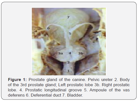
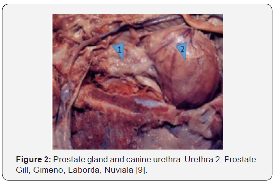
The urethra crosses the prostate before reaching the base of the penis, the vas deferens also pass through a small portion of the cranial part of the gland before penetrating dorsally into the urethra, this gland produces a transparent secretion that is expelled into the urethra [11]. The function of the prostate gland is to produce a seminal fluid called prostatic fluid, which provides an optimal environment for survival and sperm motility [12]. The prostatic fluid is milky and alkaline; This neutralizes the urethral acidity, helps the activation of sperm, gives consistency to the seminal fluid and gives the characteristic smell of semen [13]. The prostate usually covers 50% or a smaller percentage of the diameter of the pelvic inlet. The gland will be intrapelvic in young dogs and will become intra-abdominal as the prostate enlarges in older dogs [14]. Within the sexual maturity of the canine the size of this gland increases until it reaches adult size, but this growth continues to increase slowly over time, the prostate expansion is accompanied by the age of the dog, the older there is more expansion of this, which brings certain pathologies involved [9]. The physical examination of the prostate is established by abdominal and rectal palpation and the enlarged gland is rarely located completely within the pelvic canal. The palpation is done to assess the size, shape, symmetry, consistency and mobility of the prostate as well as to detect any discomfort (Figure 1 & 2) [15].
The Prostate Gland and the Pelvic Urethra
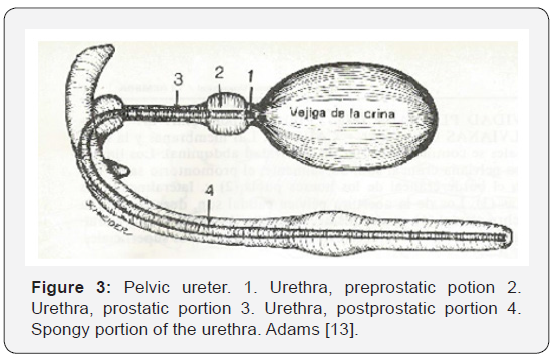
The prostate and the urethra are anatomical structures that are strongly involved, since both perform functions together. The urethra is a conduit that has the mission of transporting urine and semen to the end of the penis [11]. The canine urethra is composed of a pelvic portion inside the pelvis and another portion of the penis. The pelvic portion has pre-prostatic and prostatic components (Figure 3), [10]. The pelvic urethra in its pre-prostatic portion is short and lies between the urinary bladder and the prostate; On the other hand, the prostatic portion of the urethra is surrounded by the prostate in its entirety, while the lobes of the gland are surrounding the urethra [13]. The urethra traverses the prostate before reaching the base of the penis and the vas deferens also passes a small portion of the cranial part of the gland before penetrating dorsally into the urethra. The urethra in its pelvic portion has a beginning in the neck of the bladder and maintains a lot of proximity to the prostate gland and goes caudally on the floor of the pelvis, crossing the prostate inside thus giving shape to the longitudinal groove that divides the gland Prostatic in its two lobes [16].
Anatomical Differences of the Prostate Gland in Dogs and Cats
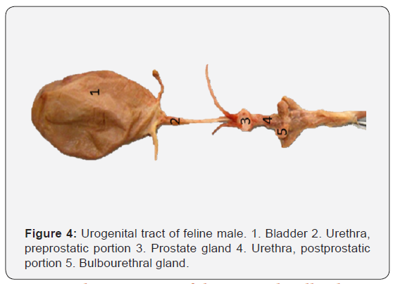
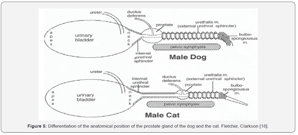
The cat and the dog share an accessory sex gland, which is the prostate gland while the prostate and the urethral bulb gland are present in the cat (Figure 4). The prostate gland has two parts: the body and the disseminated part. In the dog, the body of this gland has two spherical lobes, two cranial and two caudal, approximately 1cm in length or less, covering the dorsal urethra and laterally on the neck of the bladder [12]. In the dog, the body of the prostate is spherical and completely surrounds the urethra and the beginning of the bladder, (right and left) with an uneven surface [17]. In contrast, the cat has its prostate gland composed of four lobes (Figure 4 & 5).
Anatomical Description of the Seminal Colliculi
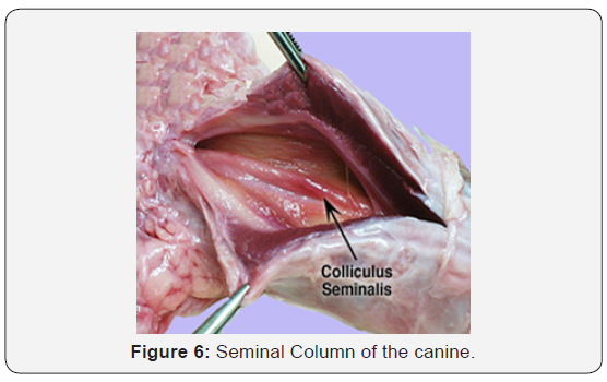
A seminal colliculus (Figure 6) is described as an elevation of the mucosa in the prostatic part of the urethra [13]. The first part of the urethra (pelvic portion) is entirely surrounded by the prostate and its light has a dorsal ridge that makes inward relief [19]. In the urethra, specifically in the dorsal or urethral crest, a protrusion or elevation of the dorsal wall of the prostatic urethra is born and, near its middle part, a dorsal crest protrudes in the urethral lumen, which continues to thicken or thicken. The seminal colliculus is perforated on each side by the narrow opening of the vas deferens and the numerous pores that drain the prostate.
The anatomical relationships of the seminal colliculus with other nearby structures (See Figure No. 3) is first given by an intimate relationship between the colliculus and the prostate, since the latter has numerous prostatic ducts, which empty their contents into the urethra. prostatic near the opening of the vas deferens, thus involving the seminal colliculus. Johnston [12]; second, the colliculus and the vas deferens are also linked, since these ducts carry sperm from the epididymis to the urethra and pass laterally to the seminal colliculus through its openings [11]. The vas deferens by their craniolateral aspect of each prostate wolf course caudoventrally before entering the urethra immediately next to the seminal colliculus (Figure 6) [20].
Conclusion
The bibliographical study carried out based on the prostate gland and the seminal colliculi is analyzed. The anatomical relationships are fundamental for the full functioning of the organism in general, since to be able to study the anatomy of the canine can be understood that fit all anatomical structures at the time of relating them to each other. The connectivity of the prostate gland and the seminal colliculi based on their union structure, which is the urethra, is fundamental for the manufacturing process, enrichment and arrival of the semen to the urethra, thus also involving the seminal colliculi.
To Know More About Anatomy Physiology & Biochemistry International Journal Please click on:
For more Open Access Journals in Juniper Publishers please click on:

Comments
Post a Comment