Cavum Septi Pellucidiet Vergae: Anatomic Finding and Review-Juniper Publishers
JUNIPER PUBLISHERS-OPEN
ACCESS ANATOMY PHYSIOLOGY & BIOCHEMISTRY INTERNATIONAL JOURNAL

The authors discuss in this article about the cavum
septi pellucidi and cavum Vergae. The septum pellucidum is a transparent
part of two laminae that extends from the anterior part of the body
(the genu) to the superior surface of the fornix. The development of the
septum pellucidum occurs from the primitive lamina terminalis which
forms the anterior wall of the telencephalic cavity. The article
presents a mini-review on the subject and it includes information about
their embryology and anatomic findings.
Keywords: Anatomy; Cavumseptumpellucidum; Cavumseptipellucidi; Cavumvergae; Verga’s Ventricle
Embryology of the Septum
Derived from the lateral part of the terminal bar,
firstly, the septum binds to the frontal direction, always separated
from the pallial part by a ventricular groove, a septum-cortical
fissure. This pre commissural part of the septum has a considerable
thickness, especially in lower mammalians. As with the reptiles, their
cells are grouped into two nuclei, the septi nuclei dorsolateral and
ventromedial. It contains the fibers that ascend to the hippocampus
(olfactory hyppocampal fibers or olfactory bundle of the septum). The
pre commissural part continues backward, below the corpus callosum and
over the foramen of Monro. This supra foraminal of the septum contains
the fibers fornicais and cortico-habenulares. The extension of the
corpus callosum allows an extension of the septum, whose pre commissural
part is reduced as the measure that increases the frontal extension of
the corpus callosum, while the supra foraminal suffers a stretching and a
reduction still more important, as a result of the posterior
displacement of the Amon horn. Such septum extension implies in its
reduction to a fine membrane, the septum pellucidi of a man. Among the
septal nucleus, the ventro medial nucleus, located in front of the
anterior commissure, only maintains a remarkable volume. The bilateral
septi can suffer coalescence or even a concrescence so that the
arachnoid space, a septum cavity located between the septi of both
sides, disappears. This process is almost normal in its posterior part.
On the other hand, an increase of the arachnoid fluid can cause an
expansion of the interseptal sub callosal space, including its posterior
part, which, then, is transformed and becomes the cavity named Verga.
However, it is evident that these cavities do not have any normal
communication with the cerebral ventricles [1-3].
Anatomy of the Septum
The septum, in the ventromedial portion of the
hemispheres of the forebrain, is differentiated in all vertebrates [4].
The septum pellucidum of man is composed of two thin leaves of gliotic
tissue with occasional scattered neurons. The leaves may be fused, but
an interseptal space, the cavum septi pellucidi, persists in 85 per cent
of normal adult as a small slit or large space [5]. This interseptal
space is lined by the ependyma [6] and it is often connected with the
ventricular system, as known by the frequency of filling with air during
pneumo encephalography, even in neonates [7]. A combination of the
theories would explain the histological nature of the septal leaves as
neural tissue lined by ependyma and derived from the lamina terminalis
the cavum septi pellucidi may be continuous with the third ventricle, or
the laminae terminalis may completely separate the cavum from the
anterior end of the ventricle: the open cavum septi pellucidi described
by Thompson [8] as occurring in ungulates, carnivorous, and primates,
was really the recess beneath the frontal end of the corpus callosum
[9]. The human septum pellucidum is absent in association with agenesis
of the corpus callosum [10] and is intimately related to and dependent
upon the proceeding and concomitant growth of the corpus callosum during
embryogenesis [11,12].
The Ventricular System
The ventricular system is constituted from the cavities of
the cerebral vesicles and the medullar tube. All the ventricular
system is intercommunicated, demonstrating its origin of a
common cavity, the neural tube cavity. The changes of the cavities
are due to an uneven growth of the vesicles and their walls. At
the level of the medullar tube, the growth is restricted and the
walls suffer a large thickening. Consequently, the cavity becomes
relatively thin and will constitute the ependymal channel. At
the level of the rhombencephalon we will have an expanded
cavity, in spite of the pronounced growth of the walls which will
constitute the floor (medulla oblongata and pons) and the roof
(cerebellum) - the 4th ventricle. The caudal portion of the roof
of the 4th ventricle is formed by the plate of the miencephalon
roof, which is stretched and consisted of few spongioblasts
layers – is the coroidal plate of the 4th ventricle. At the level of
the mesencephalon, the growth of the walls is relatively larger
than that of the cavity, becoming reduced to a channel of small
diameter - the cerebral aqueduct, which connects with the
4th and 3rd ventricles. At the level of the diencephalon it was
observed that there is a large growth of the lateral walls, forming
the thalamic region, while the cavity is reduced to a narrow
sagittal cleft – the 3rd. ventricle. Its roof remains thin with some
thick cellular layers – the coroidal layer of the 3rd ventricle.
Below the corpus callosum a thin membrane is observed named
septum pellucidum (transparent septum), located between the
corpus callosum and the trigon. It has a triangular form and in
its inner lodging a small cavity which is named (wrongly named)
the fifth Verga’s ventricle. In the telencephalon the situation is
more complex due to the appearance of the hemispheric vesicles
and the primitive cavity is unfolded into two cavities, in a wide
communication with the 3rd. Ventricle - they will be the lateral
ventricles. Later the communication will be reduced, and the
lateral ventricles start to communicate with the 3rd, through two
holes - they are the inter ventricular holes. The regions where
the hemispheric vesicles are limited with the diencephalic floor
remain thin, constituting the choroidal plates of the lateral
ventricles.
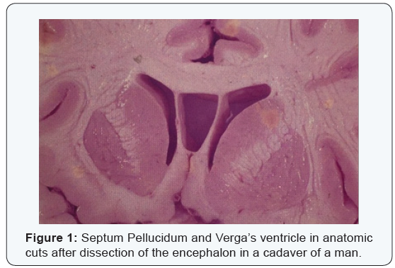
Anatomic Case
After the dissection of the encephalon in a cadaver of a man,
we observed in anatomic cuts the Verga’s ventricle (Figure 1).
The pathology and the image of the septum pellucidum and
the Verga’s ventricle are an anatomic rarity. The “cavum of the
septum pellucidum” is a cavity filled with the cerebrospinal
fluid which is situated between the frontal horns of the lateral
ventricles. The “cavum Vergae” is a posterior digiti form
extension elongated from the cavum septum pellucidum which
is located among the fornices. The cavum septum pellucidum
can occur in an isolated manner, however, the cavum Vergae only
occurs in conjunction with the cavum septum pellucidum. When
the two occur, the correct Latin nomenclature is cavum septi pellucidiet Vergae. In the daily use, the combination usually is
called cavum of the septum pellucidum [13]. The septi pellucidi
are two paired triangular membranes (“leaflets”) which develop
approximately with 12 weeks of gestational age. The embryonic
septi pellucidi are not fused, and the cavity between them is filled
with cerebrospinal fluid. This simple cavity between the two
leaflets receives two different names. Anterior to the foramen of
Monro it is called “cavum of septum pellucidum”. The posterior
continuation between the fornices is called “cavum Vergae”.
Normally, the two septi pellucidi fuse, and the cavity between
them is obliterated. The fused membranes become the septum
pellucidum. The presence of the cavum of the septum pellucidum
usually is asymptomatic and is an alteration of the type “don’t
touch”, found accidentally in the image exams. The computed
tomography and the magnetic resonance imaging of these two
entities show a cleft cavity not much visible and a prominent
collection measuring various millimeters in diameter. In rare
cases, these pathologies exceptionally increased determine
an expansive effect, displacing laterally the fornices and the
leaflets of the septi pellucidi. Those two pathologies should
not be confused with a “cavum velum enterpositum”, which
is the space of the triangular cerebrospinal fluid, thin, which
recovers the thalamus and the third ventricle. The “cavum velum
interpositum” usually occurs without the cavum septi pellucidi.
Conflict of Interest
The authors declare that they have no financial interest or
any conflicts of interest in this research.
For more Open Access Journals in Juniper Publishers please
click on: https://juniperpublishers.com
For more articles in Anatomy Physiology & Biochemistry International Journal please click on: https://juniperpublishers.com/apbij/index.php
For more Open Access Journals please click on: https://juniperpublishers.com
To know more about Juniper Publishers please click on: https://juniperpublishers.business.site/


Comments
Post a Comment