Two Cases of Inverted Ectopic Teeth in Maxillary Sinus-Juniper publishers
JUNIPER PUBLISHERS-OPEN ACCESS ANATOMY PHYSIOLOGY & BIOCHEMISTRY INTERNATIONAL JOURNAL
Asymptomatic supernumerary ectopic teeth are usually
found as incidental findings in panoramic radiographs. If present,
symptoms mostly occur according to the localization of the ectopic
teeth. Supernumerary inverted ectopic teeth in maxillary sinuses are
rare. To overcome possible complications and to assess associated
pathology, further radiographic examinations are recommended to evaluate
the exact localization of tooth and peripheral tissues. Two cases of
asymptomatic supernumerary ectopic inverted teeth are described in the
present paper. Both cases are investigated further by tomographic
examinations and are found in relation with maxillary sinus. Since the
cases were asymptomatic no surgery was performed and the patients were
called for routine follow-ups. The exact localization and peripheral
tissues of ectopic teeth should be examined carefully to overcome
possible associated pathologies and complications.
Keywords: Asymptomatic; Ectopic eruption; Inverted tooth; Maxillary sinus; Supernumerary tooth; Supplemental toothAbbreviations: CT: Computerized tomography; CBCT: Cone beam computerized tomography
Introduction
Teeth, impacted at unusual positions far from their
normal anatomical location, are called ectopic teeth [1]. An extra tooth
with an abnormal morphology is usually called as supernumerary tooth,
whereas it is called as supplementary tooth if the morphology is normal
[2,3]. The reported prevalence of supernumerary teeth varies between
%0,45 and %3 and they might erupt ectopically. There is female
predominance with a ratio of 2:1 [2]. Ectopic teeth may erupt in various
sites such as mandibular condyle and coronoid processes, maxillary
sinus, orbital floor, palate, facial skin, nasal cavity, inferior nasal
concha and oropharynx. Most supernumerary teeth are located in the
anterior maxillary region and ectopic eruption of teeth in maxillary
sinus is rare [3-6]. The present paper reports one inverted
supernumerary and one inverted supplemental ectopic tooth located in
maxillary sinuses.
Case Reports
Case 1
A 16 year old girl was referred to Istanbul
University Oral and Maxillofacial Radiology Department for orthodontic
complaints. Root fragments of 16 tooth and caries at 27,36 was seen in
intraoral examination.

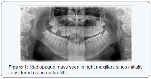
Panoramic radiography showed radiolucent lesion
located at left ramus and a radiopaque body at right maxillary sinus
(Figure 1). Radiopaque body was considered as an antrolith. Computerized
tomographic (CT) examination is performed for maxilla and mandible to
investigate the radiopaque mass in right maxillary sinus and radiolucent
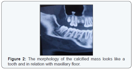
area in left mandibular ramus area. CT images showed that radiopaque
body located in right maxillary sinus has enamel like density at its
periphery and looks like a tooth crown. This body is considered as a
supernumerary
ectopic third molar because of its location and morphology
(Figure 2).

Surgery was planned for radiolucent area in left mandibular
ramus area as extraction of 38 number tooth and removal of
the radiolucent mass. Consequently there wasn’t any symptom,
sign of infection in sinus or cystic formation around impacted
tooth, surgery was not planned and patient called-up for routine
controls. During patient’s routine control session after two
years, the patient had started orthodontic theraphy and control
panoramic radiography revealed that the radiopaque body
located in right maxillary sinus is still exists and the area in left
mandibular ramus area after surgical extraction of 38 number
tooth and its associated pathology was healed (Figure 3).

Case 2

A 36 year old systemically healthy Caucasian male patient
was referred to Istanbul University Oral and Maxillofacial
Radiology Department for pain at 27 number teeth. Intraoral
examination revealed carious cavities at 14, 27, 38. Patient’s
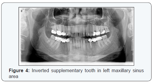
panoramic radiography showed an inverted extra tooth (Figure4). Because of its normal morphology the tooth was considered
as supplemental ectopic tooth. Deficient root canal treatment
at 15, overfilled root canal treatment at 16 teeth, apical lesions
at 25, 35 were also observed in panoramic radiography. Cone
beam computerized tomography(CBCT) was to assess the exact
location of the tooth and its relation with peripheral tissues.
Tomographic images showed that the tooth was located inside
the sinus and nasal cavity partially. Only root of the ectopic tooth
was surrounded by sinus mucosa locally and tip of the crown
was located inside nasal cavity (Figure 5). Since the patient
didn’t have any symptoms like sinusitis, foul odor, no surgery
was planned and patient was called for control examinations.

Discussion
Abnormal tissue interactions during odontogenesis
may lead to ectopic tooth development. The etiology may
include developmental pathologies such as cleft palate, fusion
deficiencies or cyst formations. Pressure by cystic enlargement
may displace the tooth buds. Genetic factors, trauma, maxillary
and odontogenic infections, high bone density, obstruction
caused by supernumerous teeth formation are also involved [6].
Teeth located at maxillary sinus may cause sinusitis or ophthalmic
symptoms but may also be remained undiagnosed for years and
can be seen as an incidental finding. The symptoms include
chronic or recurrent sinusitis, nasolacrimal duct obstruction,
nasal polyps, osteomeatal complex disease, epistaxis, facial pain,
headache, foul smell and elevation of the floor of the orbit [5-7].
Differential diagnosis should include foreign body, anthrolith,
rhinolith, sinolith, bony sequestrum, exostosis and neoplasm
[4,5]. In case 1, the first review of the panoramic radiography lead
the authors assess the mass as an antrolith. However, according
to the morphology of the mass and the peripheral enamel like
radiopacity, the mass was considered as ectopic tooth after CT
examination. Although no histological examination of the tooth
was performed, the connection of the apex dentis between the
adjacent peripheral tissues was a strong support for our claim.
Additionally the pulpal cavity at corona dentis and radical
root canal could be seen. At the coronal side, two cuspes and
considerable radiopacity at the crown which looked like enamel
tissue were observed. This situation appreciates the importance
of CT in the evaluation of masses in maxillary sinuses.
Panoramic radiography is not a suitable screening tool to
determine the periphery and exact localization of ectopic tooth
in maxillary sinus. Even though the ectopic tooth in maxillary
sinus is asymptomatic, a CBCT examination is recommended
to assess possible pathology and the certain localization of the
ectopic tooth. The patient should be recalled periodically for
evaluation of any alterations and prediction of complications.
Ectopic teeth may obstruct the osteomeatal complex of the
maxillary sinus and cause mucoceles [8]. A case of bilaterally
placed ectopic molars in the maxillary sinuses is reported by
Jude at al, in which the right ectopic molar in the maxillary right
sinus was blocking the flow of fluid, which resulted in swelling
and removed [9]. A literature review has revealed that only 30
cases have been reported in 30 years between 1980-2010 and
only 6 of the cases were asymptomatic and noticed incidentally.
Both cases in the present paper are asymptomatic [10].
For some authors, standard treatment for an ectopic tooth
is extraction [11], whilst, for others, treatment of choice in
asymptomatic ectopic tooth cases is continued observation [12].
Ectopic teeth tend to form a cyst or tumor if not managed [13].
Because both of the cases were asymptomatic, sole observation
was preferred, and the teeth were left untouched.
Conclusion
Ectopic tooth in maxillary sinus should be taken into
consideration whether it manifests symptoms or not, since it
may cause pathological alterations within time.
For
more Open Access Journals in Juniper Publishers please
click on: https://juniperpublishers.com
For more articles in Anatomy Physiology & Biochemistry International Journal please click
on: https://juniperpublishers.com/apbij/index.php
For more Open Access Journals please click on: https://juniperpublishers.com
To know more about Juniper Publishers please click on: https://juniperpublishers.business.site/


Comments
Post a Comment